Ultrasound-Guided Selective Nerve Root Block for Surgical Planning in Multilevel Cervical Disc Disease: A Technical Report
King Hei Stanley Lam, Yonghyun Yoon, Daniel Chiung-Jui Su, Anwar Suhaimi, Teinny Suryadi, Abdallah El-Sayed Allam, Manal Hassanien
Cureus 18(3): e105690
Abstract
Selecting the appropriate surgical level in patients with multilevel cervical disc disease remains challenging when relying solely on clinical examination and magnetic resonance imaging (MRI) findings. Fluoroscopy-guided selective nerve root blocks (SNRBs) carry risks of vascular injury from inadvertent intra-arterial injection. This technical report describes the technical aspects of ultrasound-guided SNRB for preoperative surgical-level localization and provides a reproducible protocol for clinical implementation.
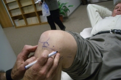
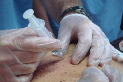
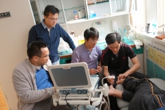
The procedure utilizes a high-frequency (12 MHz) linear transducer with color/power Doppler capability. Transverse process morphology guides level identification: C7 exhibits a rudimentary anterior tubercle with a prominent posterior tubercle; C6 displays a sharp, prominent anterior tubercle; and C3-C5 demonstrate the characteristic "two-humped camel" sign. The vertebral artery is identified anterior to C7 and confirmed with Doppler. Using an in-plane posterolateral to anteromedial approach, a 25-gauge needle is advanced toward the oval hypoechoic nerve root between the anterior and posterior tubercles (or anterior to the posterior tubercle for C7). After negative aspiration, 1 mL of 1% lidocaine is injected. For multilevel assessment, sequential blocks are performed, with a minimum of four-hour intervals between injections, blocking from caudal to cephalad. A positive response is defined as ≥50% reduction in arm pain on the Visual Analog Scale at 30 minutes post-injection.
This technique was developed and validated during a randomized controlled trial (NCT05145530), whose clinical outcomes have been published separately. The present technical report focuses on the procedural aspects. Ultrasound guidance enabled consistent visualization of target nerve roots in all 30 intervention patients (72 blocks). Power Doppler identified radicular arteries adjacent to nerve roots in 25% of procedures, allowing trajectory adjustment to avoid vascular puncture. No procedure-related complications occurred. The sequential block protocol successfully identified symptomatic levels in all patients, demonstrating the feasibility of the technique to inform surgical planning.
Ultrasound-guided cervical SNRB is a safe, radiation-free technique that enables real-time visualization of nerve roots and vascular structures, thereby enhancing the safety profile compared to fluoroscopy-guided approaches. The detailed step-by-step technique described herein should enable other clinicians to incorporate this approach into practice for accurate preoperative level selection in patients with multilevel cervical disc disease.

Annual Scientific Meeting 2026
Date: 19 - 20 September 2026 (Saturday - Sunday)
Diploma of Musculoskeletal Medicine 2025 - 2027
Registration Deadline: 31 August 2025
Enquiry: hkimmltd@gmail.com